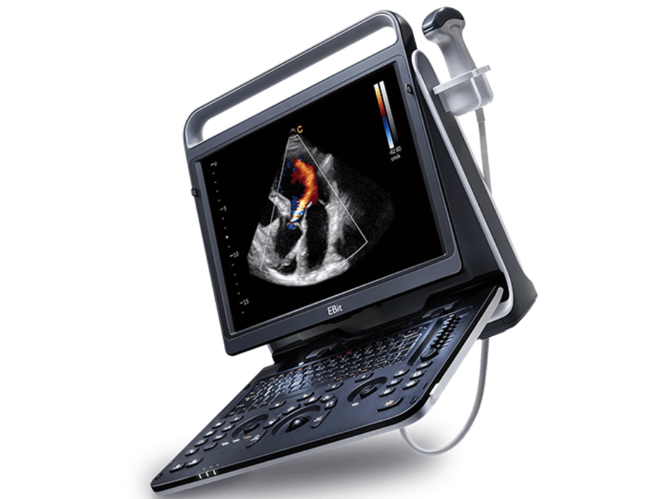
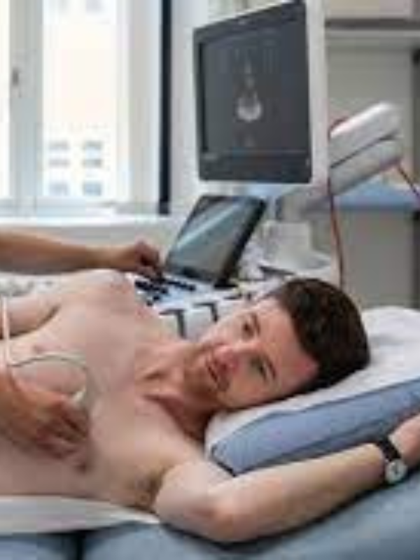
An echocardiogram, commonly known as an ECHO, is a sonographic imaging technique that uses ultrasound waves to create images of the heart. This non-invasive procedure is a key tool at Sahan Diagnostic Center for assessing the heart’s structure and function, aiding in the diagnosis and management of various cardiac conditions.
Some of the Features
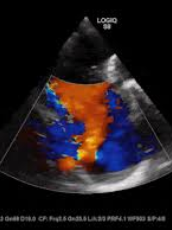
Real-Time Heart Imaging
It provides real-time images of the heart, allowing for the assessment of the heart's chambers, valves, walls, and blood vessels.
Non-Invasive and Safe
Echocardiography is a painless procedure that does not use ionizing radiation, making it safe for all patients, including those with chronic health conditions and pregnant women.
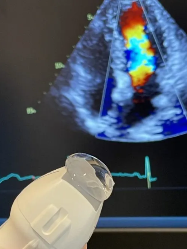
Doppler Echocardiography
This technique measures The heart's walls, which can indicate how well the heart pumps blood and can help in identifying areas of the heart muscle that aren't contracting effectively
Uses of the 128-Slice CT Scanner
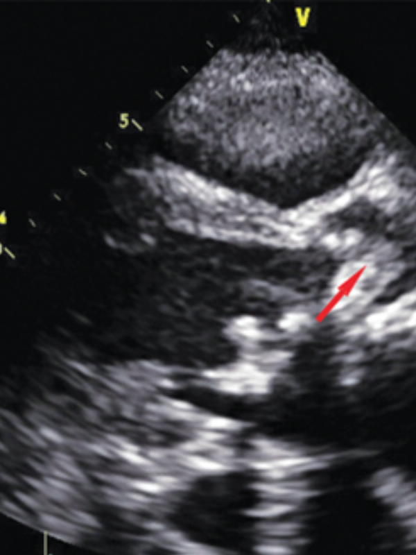
Diagnosis of Heart Conditions
Echocardiography is crucial for identifying abnormalities in heart valves, such as stenosis or regurgitation, and is essential in diagnosing various types of cardiomyopathies and congenital heart defects.
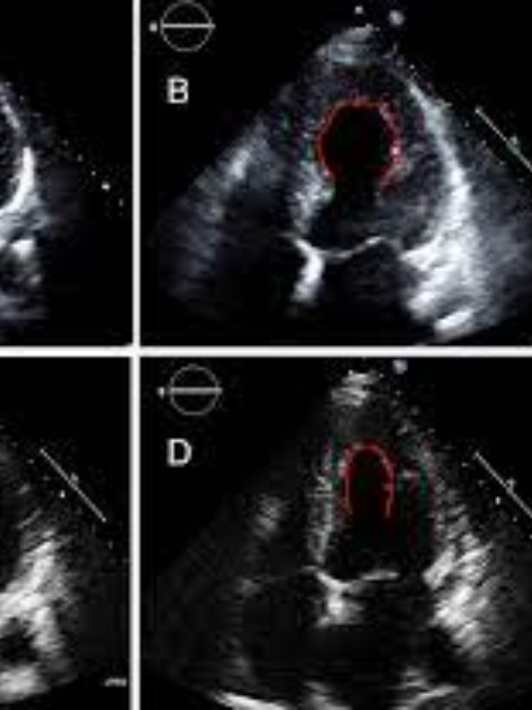
Assessing Cardiac Function
It measures the ejection fraction to gauge heart function and assesses diastolic function to understand how well the heart relaxes and fills with blood.
Evaluating Heart Failure
It helps determine the underlying cause of heart failure and monitors the effectiveness of treatments, whether due to weakened or stiff heart muscle or valvular heart disease.
Detecting Heart Muscle Damage
Post-heart attack, it can show areas of the heart muscle that are not contracting normally, and it’s also used in diagnosing myocarditis, the inflammation of the heart muscle.
Guiding Cardiac Procedures
It guides interventional procedures like balloon valvuloplasty and assists in guiding needle biopsies of the heart muscle.